File:Symptoms_of_fibromyalgia.png
From Wikipedia, the free encyclopedia
मूल चित्र ((730 × 688 पिक्सेल, फ़ाइल का आकार: 155 KB, MIME प्रकार: image/png))
|
|
यह फ़ाइल विकिमेडिया कॉमन्स से है। वहाँ पर इसका विवरण पृष्ठ निम्नोक्त है। कॉमन्स मुक्त लाइसेंसों के अंतर्गत उपलब्ध मीडिया फ़ाइलों का संग्रह है। आप भी इसमें मदद कर सकते हैं। |
सामग्री
सारांश
| विवरणSymptoms of fibromyalgia.png |
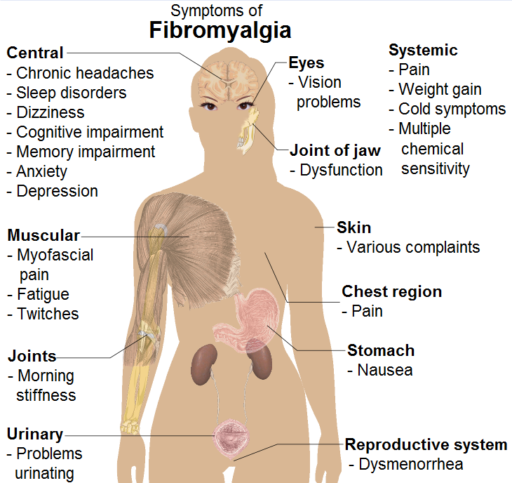
English: Common signs and symptoms of fibromyalgia. (See Wikipedia:Fibromyalgia#Signs and symptoms).
Model: Mikael Häggström. To discuss image, please see Template talk:Human body diagrams
References
|
| दिनांक | |
| स्रोत | All used images are in public domain. |
| लेखक |
When using this image in external works, it may be cited as:
or
|
| दूसरे संस्करण |
 |

|
File:Symptoms of fibromyalgia.svg is a vector version of this file. It should be used in place of this PNG file when not inferior.
File:Symptoms of fibromyalgia.png → File:Symptoms of fibromyalgia.svg
For more information, see Help:SVG.
|
 |
लाइसेंस
| Public domainPublic domainfalsefalse |
| मैं, इस कार्य का/की कॉपीराइट धारक, इस कार्य को सार्वजनिक डोमेन में प्रकाशित करता/करती हूँ। यह पूरे विश्व में लागू होता है। कुछ देशों में यह कानूनी तौर पर नहीं हो सकता है; ऐसा हो तो: मैं सभी को इस कार्य का इस्तेमाल किसी भी उद्देश्य से, बिना किसी बाधाओं के इन शर्तों के कानून द्वारा अनिवार्य किए तक करने की अनुमति देता/देती हूँ। |
Human body diagramsMain article at: Human body diagrams Template location:Template:Human body diagrams How to derive an imageDerive directly from raster image with organsThe raster (.png format) images below have most commonly used organs already included, and text and lines can be added in almost any graphics editor. This is the easiest method, but does not leave any room for customizing what organs are shown. Adding text and lines: Derive "from scratch"By this method, body diagrams can be derived by pasting organs into one of the "plain" body images shown below. This method requires a graphics editor that can handle transparent images, in order to avoid white squares around the organs when pasting onto the body image. Pictures of organs are found on the project's main page. These were originally adapted to fit the male shadow/silhouette.
Organs:
Derive by vector templateThe Vector templates below can be used to derive images with, for example, Inkscape. This is the method with the greatest potential. See Human body diagrams/Inkscape tutorial for a basic description in how to do this.
Examples of derived works
Licensing
|


.png)


Captions
Items portrayed in this file
चित्रण
१९ अप्रैल 2009
media type अंग्रेज़ी
image/png
चित्र का इतिहास
फ़ाइलका पुराना अवतरण देखने के लिये दिनांक/समय पर क्लिक करें।
| दिनांक/समय | थंबनेल | आकार | सदस्य | प्रतिक्रिया | |
|---|---|---|---|---|---|
| वर्तमान | 06:50, 12 दिसम्बर 2009 | | 730 × 688 (155 KB) | Mikael Häggström | +jaw joint |
| 15:52, 11 दिसम्बर 2009 |  | 730 × 688 (152 KB) | Mikael Häggström | Replaced myself with the female shadow, since it affects more females than males, with a ratio of approximately 9:1 | |
| 07:48, 19 अप्रैल 2009 |  | 777 × 675 (430 KB) | Mikael Häggström | spelling | |
| 07:38, 19 अप्रैल 2009 |  | 777 × 675 (430 KB) | Mikael Häggström | {{Information |Description={{en|1=g}} |Source=Own work by uploader |Author=Mikael Häggström |Date=g |Permission= |other_versions= }} <!--{{ImageUpload|full}}--> |
चित्र का उपयोग
इस चित्र से कोई पन्ने नहीं जुड़ते
चित्र का वैश्विक उपयोग
इस चित्र का उपयोग इन दूसरे विकियों में किया जाता है:
- en.wikipedia.org पर उपयोग
- en.wikiversity.org पर उपयोग
- is.wikibooks.org पर उपयोग
- it.wikipedia.org पर उपयोग
- simple.wikipedia.org पर उपयोग
मेटाडेटा
इस फ़ाइल में अतिरिक्त जानकारी मौजूद है, जो शायद इसे बनाने या डिजिटाइज़ करने के लिए उपयुक्त डिजिटल कैमरा या फिर स्कैनर द्वारा जोड़ा गया हो।
अगर चित्र को इसके मूल रूप से बदला गया है, शायद कुछ जानकारी इसके वर्तमान स्थिति से संबंधित न हो।
| क्षैतिज रेसोल्यूशन | 37.79 dpc |
|---|---|
| वर्टिकल रिज़ोल्यूशन | 37.79 dpc |