File:Human_brain_right_dissected_lateral_view_description.JPG
From Wikipedia, the free encyclopedia
Human_brain_right_dissected_lateral_view_description.JPG (653 × 413 pixels, file size: 40 KB, MIME type: image/jpeg)
| This is a file from the Wikimedia Commons. Information from its description page there is shown below. Commons is a freely licensed media file repository. You can help. |
Summary
| DescriptionHuman brain right dissected lateral view description.JPG |
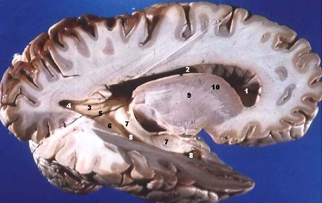
Human brain right dissected lateral view description.JPG Lateral Portion of Frontal, Parietal, Occipital, and Superior Portion of Temporal Lobe Resected. The anterior horn of the lateral ventricle is located in the frontal lobe. The body of the lateral ventricle continues posteriorly into the parietal lobe, the posterior horn into the occipital lobe, and the inferior horn down into the temporal lobe. Some structures produce elevations or bumps in the walls of the posterior and/or inferior horns of the lateral ventricles.
|
| Date | |
| Source | http://www.healcentral.org/healapp/showMetadata?metadataId=40566 (Internet Archive of file description page) |
| Author |
John A Beal, PhD Dep't. of Cellular Biology & Anatomy, Louisiana State University Health Sciences Center Shreveport |
| Permission (Reusing this file) |
CC-BY |
| Other versions | http://commons.wikimedia.org/wiki/Image:Human_brain_right_dissected_lateral_view.JPG |
Licensing
- You are free:
- to share – to copy, distribute and transmit the work
- to remix – to adapt the work
- Under the following conditions:
- attribution – You must give appropriate credit, provide a link to the license, and indicate if changes were made. You may do so in any reasonable manner, but not in any way that suggests the licensor endorses you or your use.
This file, which was originally posted to
https://web.archive.org/web/20110514023714/http://www.healcentral.org/healapp/showMetadata?metadataId=40566, was reviewed on 1 November 2013 by reviewer Avenue, who confirmed that it was available there under the stated license on that date.
|
| Annotations InfoField | This image is annotated: View the annotations at Commons |
Ventriculus lateralis, Cornu frontale
Ventriculus lateralis, Pars centralis
Calcar avis
Ventriculus lateralis, Cornu occipitale
Trigonum collaterale
Eminentia collateralis
Hippocampus
Ventriculus lateralis, Cornu temporale
Capsula interna
Nucleus caudatus
Captions
Items portrayed in this file
depicts
30 November 2005
image/jpeg
File history
Click on a date/time to view the file as it appeared at that time.
| Date/Time | Thumbnail | Dimensions | User | Comment | |
|---|---|---|---|---|---|
| current | 14:29, 22 June 2006 | | 653 × 413 (40 KB) | Patho | {{Information| |Description='''Human brain right dissected lateral view description.JPG''' Lateral Portion of Frontal, Parietal, Occipital, and Superior Portion of Temporal Lobe Resected. The anterior horn of the lateral ventricle is located in the fro |
File usage
Global file usage
The following other wikis use this file:
- Usage on ar.wikipedia.org
- Usage on bn.wikipedia.org
- Usage on bs.wikipedia.org
- Usage on da.wikipedia.org
- Usage on de.wikipedia.org
- Usage on de.wikibooks.org
- Usage on en.wikibooks.org
- Usage on en.wiktionary.org
- Usage on es.wikipedia.org
- Usage on eu.wikipedia.org
- Usage on fa.wikipedia.org
- Usage on fr.wikipedia.org
- Usage on fr.wikibooks.org
- Usage on gl.wiktionary.org
- Usage on he.wikipedia.org
- Usage on he.wiktionary.org
- Usage on hy.wikipedia.org
- Usage on it.wikipedia.org
- Usage on ja.wikipedia.org
- Usage on ko.wikipedia.org
- Usage on pl.wikipedia.org
- Usage on ru.wikipedia.org
View more global usage of this file.
Metadata
This file contains additional information, probably added from the digital camera or scanner used to create or digitize it.
If the file has been modified from its original state, some details may not fully reflect the modified file.
| _error | 0 |
|---|