File:Gray547.png
From Wikipedia, the free encyclopedia
Gray547.png (600 × 583 pixels, file size: 94 KB, MIME type: image/png)
| This is a file from the Wikimedia Commons. Information from its description page there is shown below. Commons is a freely licensed media file repository. You can help. |
Summary
| DescriptionGray547.png |
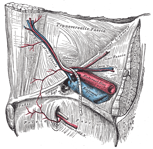
Deutsch: Blick von innen auf die Leistengegend der rechten Seite mit Darstellung der aus- und eintretenden Gefäße und anderen Strukturen. |
||||||||||||||||||||
| Plate InfoField | 547 | ||||||||||||||||||||
| Date |
before 1858 date QS:P,+1858-00-00T00:00:00Z/7,P1326,+1858-00-00T00:00:00Z/9 |
||||||||||||||||||||
| Source |
|
||||||||||||||||||||
| Author |
creator QS:P170,Q955620 |
||||||||||||||||||||
.jpg)
Book
| Henry Gray: Gray's Anatomy (20th edition)
|
|||||||||||||||||||||||
|---|---|---|---|---|---|---|---|---|---|---|---|---|---|---|---|---|---|---|---|---|---|---|---|
| Author |
|
-_Title_page.png) | |||||||||||||||||||||
| Editor |
Revised by Warren H. Lewis |
||||||||||||||||||||||
| Illustrator |
|
||||||||||||||||||||||
| Title | |||||||||||||||||||||||
| Edition |
20 |
||||||||||||||||||||||
| Publisher | |||||||||||||||||||||||
| Object type |
version, edition or translation |
||||||||||||||||||||||
| Page overview | list of all the plates | ||||||||||||||||||||||
| Language |
English |
||||||||||||||||||||||
| Publication date |
1918 |
||||||||||||||||||||||
| Place of publication |
Philadelphia / New York City |
||||||||||||||||||||||
| Source | Bartleby | ||||||||||||||||||||||

Licensing
This image is in the public domain because it is a mere mechanical scan or photocopy of a public domain original, or – from the available evidence – is so similar to such a scan or photocopy that no copyright protection can be expected to arise. The original itself is in the public domain for the following reason:
This tag is designed for use where there may be a need to assert that any enhancements (eg brightness, contrast, colour-matching, sharpening) are in themselves insufficiently creative to generate a new copyright. It can be used where it is unknown whether any enhancements have been made, as well as when the enhancements are clear but insufficient. For known raw unenhanced scans you can use an appropriate {{PD-old}} tag instead. For usage, see Commons:When to use the PD-scan tag.  | |||||
Captions
Items portrayed in this file
depicts
File history
Click on a date/time to view the file as it appeared at that time.
| Date/Time | Thumbnail | Dimensions | User | Comment | |
|---|---|---|---|---|---|
| current | 20:22, 23 January 2007 | | 600 × 583 (94 KB) | Pngbot | optimized with optipng |
| 13:30, 7 February 2006 |  | 600 × 583 (152 KB) | Arcadian | {{Gray's Anatomy plate}} |
File usage
- External iliac artery
- External iliac vein
- Femoral ring
- Inferior epigastric artery
- Inguinal canal
- Inguinal ligament
- Lacunar ligament
- Obturator artery
- Obturator canal
- Obturator foramen
- Obturator nerve
- Obturator veins
- Pelvic fascia
- Transversalis fascia
- Vas deferens
- Talk:Iliacus muscle
- User:Was a bee/Gray
Global file usage
The following other wikis use this file:
- Usage on ar.wikipedia.org
- Usage on az.wikipedia.org
- Usage on bg.wikipedia.org
- Usage on bn.wikipedia.org
- Usage on bs.wikipedia.org
- Usage on ca.wikipedia.org
- Usage on cs.wikipedia.org
- Usage on de.wikipedia.org
- Usage on de.wikibooks.org
- Usage on es.wikipedia.org
- Usage on eu.wikipedia.org
- Usage on fa.wikipedia.org
- Usage on fr.wikipedia.org
- Usage on he.wikipedia.org
- Usage on it.wikipedia.org
- Usage on ja.wikipedia.org
- Usage on ko.wikipedia.org
View more global usage of this file.